A) A brightness mode (b-mode) image of the lateral abdominal wall.

Download scientific diagram | (A) A brightness mode (b-mode) image of the lateral abdominal wall. Abbreviations: EO, external oblique; IO, internal oblique; TrA, transversus abdominis. (B) A split-screen image with b-mode on the left and motion mode (m-mode) on the right. The m-mode image represents the information from the dotted line on the b-mode image displayed over time (x-axis). Static structures produce straight interfaces while structures that change in thickness or depth (in this case the TrA) create curved interfaces. The increase in depth of the TrA correlates to a contraction. Reproduced with permission Whittaker 2007. 142 from publication: Rehabilitative Ultrasound Imaging: Understanding the Technology and Its Applications | The use of ultrasound imaging by physical therapists is growing in popularity. This commentary has 2 aims. The first is to introduce the concept of rehabilitative ultrasound imaging (RUSI), provide a definition of the scope of this emerging tool in regard to the physical | Rehabilitation, Ultrasonography and Ultrasound Imaging | ResearchGate, the professional network for scientists.

A) A brightness mode (b-mode) image of the lateral abdominal wall.

Ultrasound image of the zone of apposition of the diaphragm. In

Ultrasound Modes – basic concepts in ultrasound physics

Sonography Assessment: Overview of AFAST and TFAST
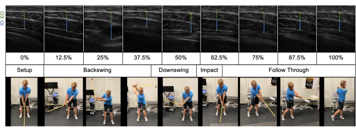
Providing Visual Biofeedback Using Brightness Mode Ultrasound During a Golf Swing

Jackie WHITTAKER, Professor (Associate), BScPT, PhD

Comparison of A-mode and B-mode Ultrasound for Measurement of Subcutaneous Fat - ScienceDirect

B-mode ultrasound using linear probe showing anterior abdominal wall

a Probe position for B and M mode diaphragmatic excursion measurements

A) A brightness mode (b-mode) image of the lateral abdominal wall.
Frontiers Preclinical Ultrasound Imaging—A Review of Techniques and Imaging Applications
Point-of-Care Ultrasound for Outpatient Neurology - Practical Neurology
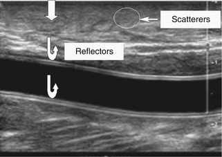
Physics and Instrumentation in Doppler and B-mode Ultrasonography