Figure 6 from Femoral Hernia: A Review of the Clinical Anatomy and

Figure 6. Femoral hernia repair in clean operation. (a) The narrow side of the mesh is sutured to Cooper’s ligament; (b) The mesh is sutured to the iliopubic tract or shelving portion of the inguinal ligament; (c) The posterior wall of the inguinal canal is reinforced, as in Lichtenstein’s repair. - "Femoral Hernia: A Review of the Clinical Anatomy and Surgical Treatment"
Fascinating history of groin hernias: Comprehensive recognition of anatomy, classic considerations for herniorrhaphy, and current controversies in hernioplasty
Cureus, Femoral Hernia Containing a Strangulated Appendix: A Hybrid Approach
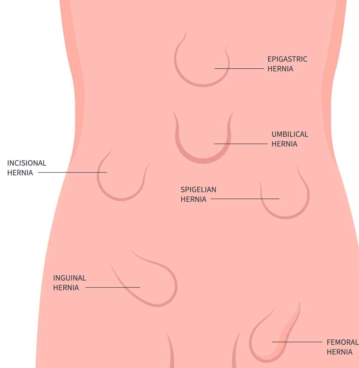
Abdominal Hernia - Epigastric - Spigelian - Obturator - TeachMeSurgery

Figure 7 from Femoral Hernia: A Review of the Clinical Anatomy and Surgical Treatment

Adult groin hernias - ScienceDirect

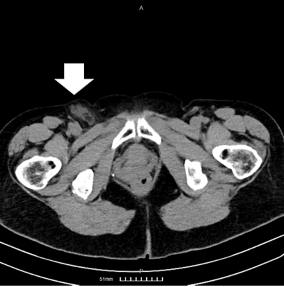
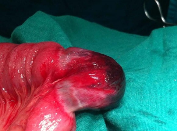
Right incarcerated femoral hernia; the contents of the hernia were the
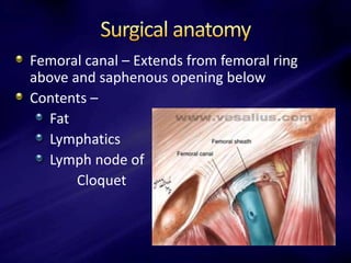
Femoral hernia anatomy
Femoral hernia
Femoral Hernia - Risk Factors - Clinical Features - Management - TeachMeSurgery

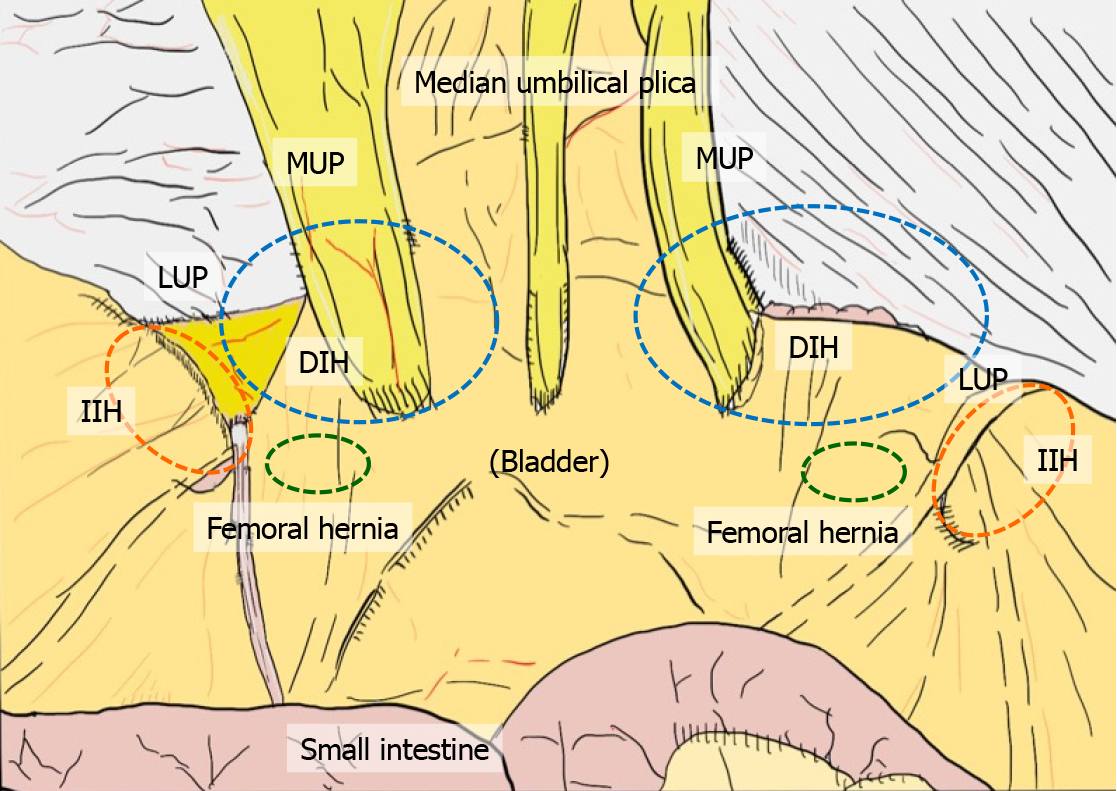
The anatomical locations of the groin hernia defects. 1: Lateral