Figure 3 from Relevant surgical anatomy of the chest wall.

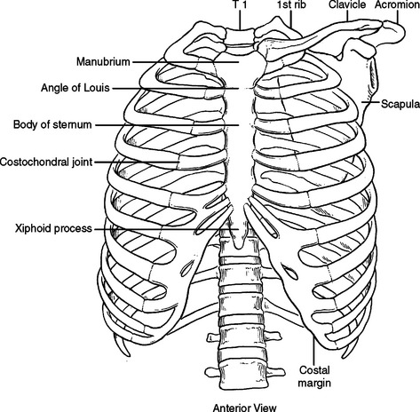
Fig. 3. Anterior chest wall showing the sternum. Note where the costal cartilages articulate with the sternum. In the intercostal space lie different structures: several kinds of intercostal muscles, intercostal arteries and associated veins, lymphatics, and nerves. (From Rendina EA, Ciccone AM. The intercostal space. Thorac Surg Clin 2007;17(4):491e501; with permission.) - "Relevant surgical anatomy of the chest wall."
PERTINENT SURGICAL ANATOMY OF THE THORAX AND MEDIASTINUM
Applied Anatomy of the Chest Wall and Mediastinum
UCSF Ortho Anatomy Core UCSF Department of Orthopaedic Surgery

SURGICAL ANATOMY OF THE CHEST WALL

Minimally Invasive Thoracic Surgery: When It's Appropriate and When It's Not

Introduction to chest wall reconstruction: anatomy and physiology of the chest and indications for chest wall reconstruction. - Abstract - Europe PMC

SURGICAL ANATOMY OF THE CHEST WALL
3: The Thorax Pocket Dentistry
/jcm/jcm-11-05516/article_deploy/html/

Musculoskeletal Imaging of Chest Wall Injuries in Athletes - ARRS InPractice

Disorders of the Chest Wall - TeachMeSurgery