LMs of living cells showing chloroplasts. Fig. 1. Girdle view of
![](https://www.researchgate.net/profile/Tamotsu-Nagumo/publication/232896141/figure/fig2/AS:328258789888001@1455274402667/3-LMs-of-living-cells-showing-chloroplasts-Fig-1-Girdle-view-of-single-cell-showing_Q320.jpg)
![](https://ars.els-cdn.com/content/image/3-s2.0-B9780323999311000714-f00071-03-9780323999311.jpg)
Frustule - an overview
![](https://static.cambridge.org/binary/version/id/urn:cambridge.org:id:binary:20200218084023584-0883:S002428291900046X:S002428291900046X_fig6.png?pub-status=live)
Symbiont composition of the basidiolichen Lichenomphalia
![](https://www.studyadda.com/upload/html_folder/6_Tissues_S-9_NZ/6_Tissues_S-9_NZ_files/image002.png)
JEE Main, JEE Advanced, CBSE, NEET, IIT, free study packages, test
![](https://www.researchgate.net/profile/Rocio-Rodiles-Hernandez/publication/258915159/figure/tbl1/AS:668430751498243@1536377718135/Counts-of-caudal-and-precaudals-vertebrae-of-Ictalurus-furcatus-I-meridionalis-and-I_Q320.jpg)
The Walter Reed Visual Assessment Scale (used with permission from
![](https://slideplayer.com/8/2304165/big_thumb.jpg)
A Tour of the Cell Chapter 6 Lecture modified by Bronwyn Scott
![](https://media.springernature.com/full/springer-static/image/art%3A10.1038%2Fs41598-017-06938-x/MediaObjects/41598_2017_6938_Fig1_HTML.jpg)
Fungal root symbionts of high-altitude vascular plants in the
![](https://img.brainkart.com/imagebk28/LdDI7os.jpg)
Living Cell
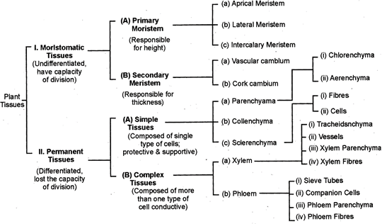
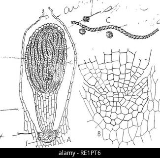
Draw a neat and labelled diagram of a sectional view of
![](https://www.researchgate.net/profile/Tsuyoshi-Watanabe-2/publication/262872516/figure/fig1/AS:392443305447424@1470577183174/LM-observations-of-cleaned-valves-in-clones-of-Surirella-cf-fastuosa-a-b-Before_Q320.jpg)
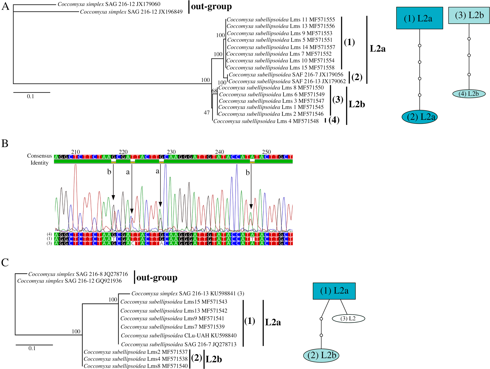
Masahiko Idei's research works Bunkyo University, Tokyo and
![](https://www.researchgate.net/profile/Sascha-Seifert-2/publication/233983279/figure/fig3/AS:339609662836739@1457980661428/Four-examples-from-the-MR-data-with-detection-results-Although-the-volunteer-in-the_Q320.jpg)
LMs of living cells showing chloroplasts. Fig. 1. Girdle view of
![](https://media.springernature.com/m685/springer-static/image/art%3A10.1007%2Fs11284-010-0712-4/MediaObjects/11284_2010_712_Fig3_HTML.gif)
A review of light interception in plant stands from leaf to canopy
![](https://www.flexiprep.com/NIOS-Notes/Senior-Secondary/Biology/posts/NIOS-Biology-Ch-4-Cell-Structure-and-Function-Part-3/Image-showing-Structure-of-a-chloroplast.png)
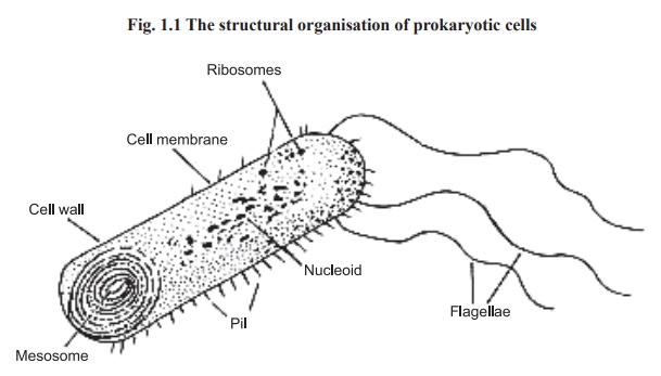
Cell Structure and Function: The Cytoplasm, the Cell Organelles and
![](https://l450v.alamy.com/450v/re1pt6/nature-and-development-of-plants-botany-development-of-plants-287-while-others-known-as-elaters-elongate-greatly-and-serve-to-con-duct-the-foods-absorbed-by-the-foot-to-the-spore-mother-cells-and-finally-they-become-spirally-thickened-fig-194-c-these-elaters-arise-through-the-sterilization-of-certain-of-the-spore-lv-pxltf-aft-fig-194-structure-of-the-nearly-mature-sporophyte-a-continuation-of-the-growth-in-fig-193-c-showing-the-formation-of-a-foot-stalk-and-capsules-c-which-contain-elongated-dark-cells-the-elaters-and-the-spores-the-archegonium-has-bee-re1pt6.jpg)
Nature and development of plants . Fig. 5. Greatly enlarged view