A 60-year-old patient presented by a lump in the left breast.
![](https://www.researchgate.net/publication/343459935/figure/fig4/AS:933529013714953@1599582069771/A-60-year-old-patient-presented-by-a-lump-in-the-left-breast-Mammography-revealed-focal.png)
![](https://www.oatext.com/img/CCRR-9-520-g001.gif)
Imaging features of acellular dermal matrix in the reconstructed
![](https://i1.rgstatic.net/ii/profile.image/11431281206690171-1700849818697_Q64/Jakub-Kufel.jpg)
PDF) Comparative study between contrast-enhanced mammography
![](https://www.researchgate.net/publication/339283419/figure/fig4/AS:858669075922944@1581734069602/A-36-year-old-woman-complains-of-a-right-breast-lump-a-Craniocaudal-and-b-mediolateral.png)
A 36-year-old woman complains of a right breast lump. a
![](https://upload.wikimedia.org/wikipedia/commons/thumb/0/03/Breast_Cancer.png/640px-Breast_Cancer.png)
Breast cancer - Wikipedia
![](https://www.independentnurse.co.uk/media/1h0ohhez/clinical-images-1.gif?width=1002&height=668&bgcolor=White&rnd=133205164658070000)
Breast cancer in images
![](https://i1.rgstatic.net/ii/profile.image/11431281119196192-1676032568787_Q64/Samantha_Gauci2.jpg)
PDF) Comparative study between contrast-enhanced mammography
![](https://www.draliabadi.com/wp-content/uploads/2015/05/Menopause-BreastChanges.jpg)
Breast Changes and Menopause Best OBGYN Los Angeles, Dr
![](https://media.springernature.com/m685/springer-static/image/art%3A10.1186%2Fs43055-020-00209-y/MediaObjects/43055_2020_209_Fig7_HTML.png)
Breast lesions in women under 25 years: radiologic-pathologic
![](https://i1.rgstatic.net/ii/profile.image/1183683818061824-1659223622405_Q64/Andrea-Cozzi-3.jpg)
PDF) Comparative study between contrast-enhanced mammography
![](https://www.researchgate.net/publication/338280648/figure/fig1/AS:842226154344448@1577813771372/Comparison-of-diagnostic-accuracy-when-mastocheck-alone-mammography-mmg-alone-and.png)
Comparison of diagnostic accuracy when mastocheck alone
![](https://www.researchgate.net/publication/303852291/figure/fig1/AS:485793652318208@1492833640516/An-illustrative-example-showing-segmentation-of-microcalcifications-and-breast-mass-in-a.png)
An illustrative example showing segmentation of microcalcifications and
![](https://www.nejm.org/na101/home/literatum/publisher/mms/journals/content/nejm/2020/nejm_2020.382.issue-9/nejmcpc1913470/20200219/images/img_medium/nejmcpc1913470_f1.jpeg)
Case 7-2020: A 52-Year-Old Man with a Mass in the Left Breast
![](https://www.researchgate.net/profile/Lokesha-Weerasinghe/publication/362098583/figure/fig1/AS:11431281153308964@1682416587402/The-high-level-architecture-of-the-application_Q320.jpg)
Comparison of diagnostic accuracy when mastocheck alone
![](https://media.springernature.com/m685/springer-static/image/art%3A10.1186%2Fs43055-021-00449-6/MediaObjects/43055_2021_449_Fig1_HTML.png)
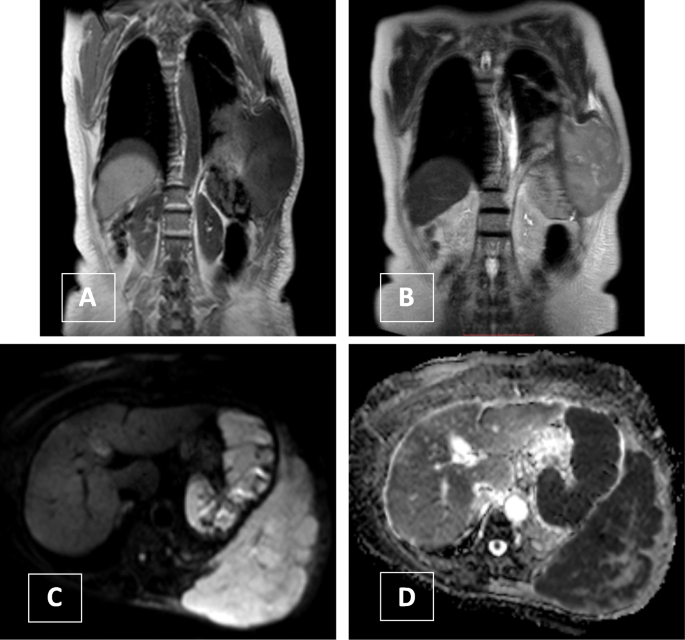
The role of MRI in comparison between benign and malignant chest wall masses in correlation with pathology, Egyptian Journal of Radiology and Nuclear Medicine