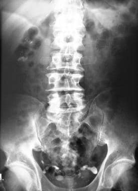
Plain X ray of a young girl with two stones in situ. (a). Stone in


Urolithiasis SpringerLink

Mukesh SURYA, Associate Professor, Indira Gandhi Medical College, Shimla, Department of Radiology

Mukesh SURYA, Associate Professor, Indira Gandhi Medical College, Shimla, Department of Radiology

Mukesh SURYA, Associate Professor, Indira Gandhi Medical College, Shimla, Department of Radiology
Cystitis Imaging: Practice Essentials, Radiography, Computed

NCCT Neck showed homogenous radio opaque mass in left tonsillar fossa.

Anjali SONI, Parul Universiy, Vadodara, Faculty of Management Studies
Plain x-ray KUB showing a single stone on the right side (white

Mukesh SURYA, Associate Professor, Indira Gandhi Medical College, Shimla, Department of Radiology

a Abdominal X-rays. a Magnet fixed with a string at the distal

Molecules, Free Full-Text

Manupriya SHARMA, Assistant Professor, MD (Pathology), DNB (Pathology), Dr Rajendra Prasad Government Medical College, Tāndā, Department of Pathology

Stonehenge - Wikipedia
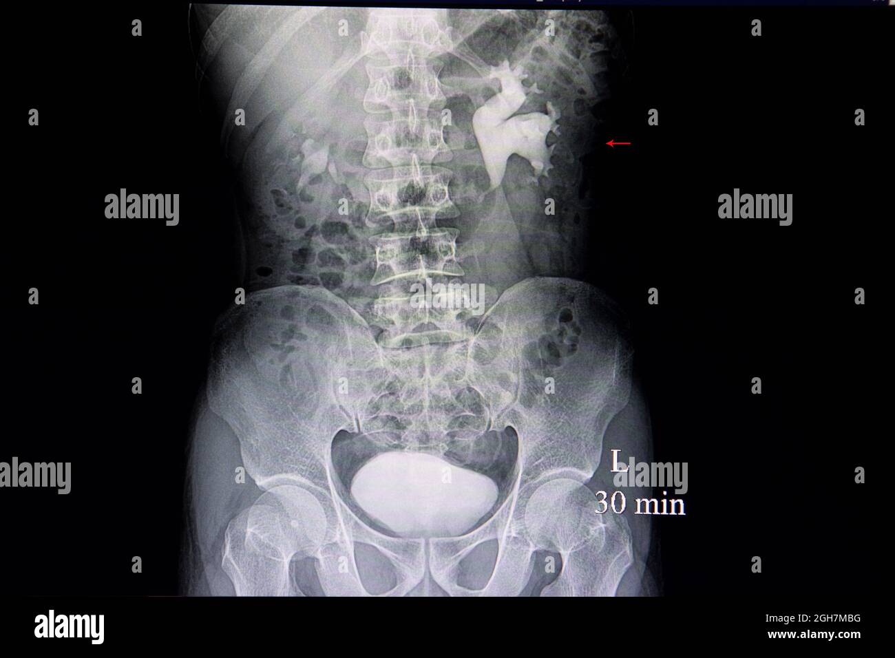
Intravenous pyelogram xray film of a patient with hydronephrosis

Xray Film Patient Large Stone Left Stock Photo 1855728682